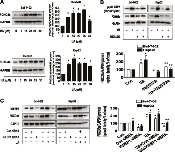
Fig. 4.

UA increased FOXO3a protein expression through activation of p38 MAPK and expression of IGFBP1. a, Bel-7402 and HepG2 cells were exposed to increased concentrations of UA for 24 h. Afterwards, the expression of FOXO3a protein was detected by Western blot. b, Bel-7402 and HepG2 cells were treated with SB203580 (10 μM) for 2 h before exposure of the cells to UA (25 μM) for an additional 24 h. Afterwards, the expression of FOXO3a protein and phoisphorylation of p38 MAPK were detected by Western blot. c, Bel-7402 and HepG2 cells were transfected with control or IGFBP1 siRNAs (50 nM each) for 24 h prior to exposure of the cells to UA (25 μM) for an additional 24 h. Afterwards, FOXO3a and IGFBP1 protein expressions were determined by Western blot, The bars represent the mean ± SD of at least three independent experiments for each condition. *Indicates significant difference as compared to the untreated control group (P < 0.05); **Indicates significance of combination treatment as compared with UA alone (P < 0.05)
