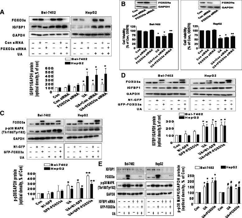
Fig. 5.

Silencing of FOXO3a overcame UA-induced cell growth inhibition and exogenous expressed FOXO3a enhanced UA-induced phosphorylation of p38 MAPK through IGFBP1. a, Bel-7402 and HepG2 cells were transfected with control and FOXO3a siRNAs for 24 h before exposing the cells to UA (25 μM) for an additional 24 h. Afterwards, FOXO3a and IGFBP1 protein expressions were determined by Western blot. b, Bel-7402 and HepG2 cells were transfected with control or FOXO3a siRNAs (up to 50 nM each) for 24 h prior to exposure of the cells to UA (25 μM) for an additional 24 h. Afterwards, FOXO3a protein expression and cell viability were determined by Western blot and MTT assays. Insert represents the protein expression of FOXO3a. c-d, Bel-7402 and HepG2 cells were transfected with control and FOXO3a overexpression vectors for 24 h before exposing the cells to UA (25 μM) for an additional 2 and 24 h, respectively. Afterwards, the protein levels of FOXO3a and p-p38 MAPK, and IGFBP1 protein expression were examined by Western blot. e, Bel-7402 and HepG2 cells silenced of IGFBP1 by siRNA previously were transfected with control and FOXO3a overexpression vector for 24 h before exposing the cells to UA (25 μM) for an additional 2 and 24 h, respectively. Afterwards, IGFBP1, FOXO3a protein and phosphorylation of p38 MAPK were determined by Western blot. Values in bar graphs were given as the mean ± SD from three independent experiments performed in triplicate. *Indicates significant difference as compared to the untreated control group (P < 0.05). **Indicates significant difference from UA treated alone (P < 0.01). #Indicates significant difference as compared to the IGFBP1 siRNA alone group (P < 0.05)
