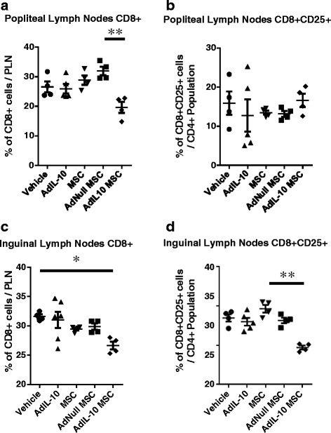
Fig. 3.

AdIL-10-transduced MSCs reduce the number of CD8 T cells in the popliteal and inguinal lymph nodes 6 weeks after injection. CD8 and CD25 expression by lymphocytes isolated from the popliteal and inguinal lymph nodes, 6 weeks post treatment. a, b AdIL-10-transduced MSCs reduced the amount of CD8+ T cells compared with AdNull-transduced MSCs in the popliteal lymph nodes, whereas no significant difference in the amount of activated CD8+ T cells was observed. c, d AdIL-10-transduced MSCs significantly decreased the amount of CD8+ cells in the inguinal lymph nodes and reduced the amount of activated CD8+ T cells compared with MSCs alone. Data points represent n = 4, pooled from eight animals. Data points in the AdIL-10 group represent n = 5, with three samples pooled from two animals and two single samples. Statistical significance was determined using a Kruskal–Wallis test, followed by Dunn’s multiple comparisons test. *p < 0.05, **p < 0.005, ***p < 0.001. Lines indicate significant difference between the two groups. ILN inguinal lymph node, MSC mesenchymal stem cell, PLN popliteal lymph node
