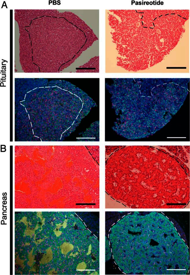
Figure 5.

Assessment by immunohistochemical analysis of effects of pasireotide on proliferation in Men1+/− mice pituitary NETs and pancreatic NETs. Pituitary and pancreatic NETs obtained from control PBS-treated Men1+/− mice are compared with pasireotide-treated Men1+/− mice, after administration of BrdU for 4 wk. A, Pituitary NET-adjacent sections from Men1+/− mice stained with H&E and immunostained for prolactin (green) and BrdU (red). Nuclei were counterstained with DAPI (Blue). The NETs are circled by dashed lines (black in the H&E sections and white in the fluorescent immunohistochemical sections). The large pituitary NETs from a PBS-treated Men1+/− mouse and a pasireotide-treated Men1+/− mouse showing immunostaining for prolactin but not GH (data not shown), are highly proliferating (red nuclei), although there are fewer proliferating cells in the pituitary NETs of the pasireotide-treated mouse. B, Pancreatic NET-adjacent sections from Men1+/− mice stained with H&E and immunostained for insulin (green) and BrdU (red), and counterstained for nuclei (blue). The NETs are circled by dashed lines (black in the H&E sections and white in the fluorescent immunohistochemical sections). The pancreatic NET from the pasireotide-treated Men1+/− mouse had fewer proliferating cells. Scale bar = 100 μm.
