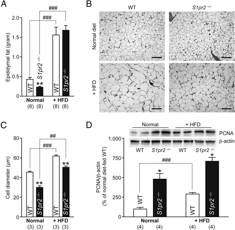
Figure 2.
HFD-induced adipocyte hyperplasia/proliferation in S1pr2−/− mice. Ten-week-old WT and S1pr2−/− mice were fed a HFD for 4 weeks. A, Epididymal fat tissue weights of normal diet- and HFD-fed mice. B, Hematoxylin/eosin-stained epididymal fat tissue sections. Scale bar, 50 μm. C, Average diameters of 300 epididymal adipocytes in each mouse line. D, Western blot analysis of PCNA expression in epididymal fat tissues. Representative blot images (duplicate) are shown as insets, and PCNA expression was quantified by normalization with β-actin levels. Data are mean ± SEM (n, sample numbers); **, P < .01 and ***, P < .001 vs WT samples; and ##, P < .01 and ###, P < .001 vs normal diet-fed samples (t test).

