Figure 5.
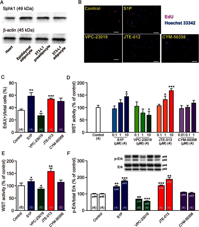
S1P and S1pr antagonists regulate preadipocyte proliferation. A, Western blot analysis of Sphk1 expression in the heart, epididymal adipocytes, and preadipocytic/differentiated 3T3-L1 cells. β-Actin was detected as loading controls. B, Representative fluorescent images of EdU (red)-labeled and Hoechst 33342 (blue)-labeled 3T3-L1 preadipocytes after a 24-hour incubation with S1P (10μM), VPC-23019 (10μM), JTE-013 (10μM), or CYM-50358 (1μM). Scale bar, 100 μm. C, Percentages of EdU-labeled (proliferative) cells/Hoechst 33342-labeled (total) cells. Over 100 Hoechst 33342-labeled cells were counted in 4 independent wells. D, WST proliferation assay of 3T3-L1 adipocytes after a 24-hour incubation with graded concentrations of S1P, VPC-23019, JTE-013, or CYM-50358. E, WST proliferation assay of 3T3-F442A adipocytes after a 24-hour incubation with S1P (10μM), VPC-23019 (10μM), JTE-013 (10μM), or CYM-50358 (1μM). F, Western blot analyses of 3T3-L1 preadipocytes using anti-Erk and anti-p-ERK antibodies. Cells were incubated with S1P (10μM), VPC-23019 (10μM), JTE-013 (10μM), or CYM-50358 (1μM) for 1 hour, and cell lysates were analyzed for Erk phosphorylation/activation. Representative images are presented. Data are mean ± SEM (n, sample numbers); *, P < .05; **, P < .01; and ***, P < .001 vs control samples (t test).

