Figure 2.
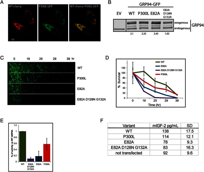
P300L GRP94 is properly expressed but less effective than WT GRP94 in supporting the production of IGF-2. A, Fluorescence microscopy analysis of expression and intracellular localization of GRP94 in grp94−/− cells. WT GRP94 mCherry-tagged was coexpressed with P300L GRP94 mGFP-tagged. Cells were fixed after 24 hours and imaged with a ×60 objective (n = 2). Bar, 5 μm. B, Comparative expression of GFP-tagged WT GRP94 or P300L, E82A, or E82A/D128N/G132A mutants of GRP94. Human embryonic kidney-293T cells were harvested 24 hours after transfection with plasmids coding for empty vector (EV) or the indicated GRP94 constructs, and protein extracts were analyzed by immunoblotting with the anti-GRP94 antibody. The lanes were run on the same gel but were noncontiguous. Quantitation of expression levels of exogenous GRP94 as compared with endogenous GRP94 is provided below lanes (n = 2). C–E, Differential ability of GRP94 mutants to support IGF-2-mediated cell survival. Grp94−/− MEFs were transiently transfected with a plasmid expressing GFP-tagged WT, P300L, E82A, or E82A/D128N/G132A variants of GRP94. Twenty-eight hours later, cell cultures were serum starved and scored at the indicated intervals by microscopy. C, Pictures of GRP94-expressing cells acquired at the indicated time points from a representative experiment. D, The number of GFP-positive cells was counted at given time points from 15 separate fields. The cell counts were plotted as a percentage of the initial time point (representative experiment shown). E, Activity of GRP94 was calculated as the survival index of cells expressing GRP94 mutants. The index was calculated at the time point within each experiment in which survival of the cells transfected with E82A/D128N/G132A GRP94 mutant was one-tenth the value of the WT-transfected cells (WT, E82A, E82A/D128N/G132A, n = 8, P300L, n = 5). Means and SDs are graphed. F, ELISA measurements of IGF-2 in supernatants of grp94−/− cells 16 hours after serum withdrawal. Grp94−/− MEFs were prepared as in panel C and secreted the indicated levels of IGF-2 in the absence of serum. The detection limit of this assay is approximately 60 pg/mL. Means and SDs are shown for two independent experiments.

