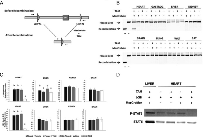
Figure 1.
Verification of inducible GHR gene disruption. A, Diagram showing exon 4 of GHR flanked by LoxP sites before and after recombination when in the presence of both MerCreMer expression and tamoxifen. Arrows denote primer positions used to distinguish floxed locus from recombined locus. B, PCR results of the floxed GHR exon 4 locus using genomic DNA isolated from eight different tissues (heart, gastrocnemius, liver, kidney, brain, lung, epididymal WAT, and brown adipose tissue) in GHRfl/fl mice without or without MerCreMer expression and with or without tamoxifen injection. C, RTqPCR analysis of GHR in heart, liver, kidney, and whole brain at 4.5 and 12.5 months of age. D, Bovine GH-stimulated STAT5 phosphorylation in 12.5-month-old heart from GHRfl/fl mice without or without MerCreMer expression and with or without tamoxifen injection and in liver from Myh6-MerCreMer+/−/GHRfl/fl (iC-GHRKO) mice. Different letters denote a difference between groups of mice (P < .05). BAT, brown adipose tissue; EX4, exon 4 of GHR; TAM, tamoxifen.

