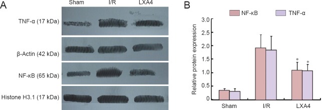
Figure 2.
Effect of lipoxin A4 (LXA4) on expression levels of TNF-α and NF-κB in the cerebral cortex of diabetic rats with cerebral ischemia/reperfusion (I/R) injury (western blot assay).
Beta-actin and Histone H3.1 were used as internal controls. The results for TNF-α were expressed as the optical density ratio of TNF-α to β-actin, and the results for NF-κB were expressed as the ratio of the optical density of NF-κB to Histone H3.1. Each bar represents the mean ± SEM (n = 6 rats per group). The data of different groups were compared by one-way analysis of variance (ANOVA) followed by Student-Newman-Keuls tests (for normally distributed data). *P < 0.05, vs. I/R. S: Sham group; I/R: diabetes mellitus + I/R injury group; LXA4: diabetes mellitus + I/R injury + lipoxin A4 group.

