
Figure 4.
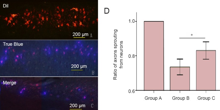
Effect of different types of Y-tube conduits injected with hWJMSCs on neuronal sprouting in the spinal cord of rats with a femoral nerve defect.
(A–C) Under a fluorescence microscope, red, blue and purple neurons are observed in the spinal cord of rats from groups A, B, and C. In rats from group A, neurons in the ventral horn of the spinal cord are stained with the red fluorescent marker Dil, indicating that axons sprouting from neurons only entered into the muscle branch of the femoral nerve. In rats from groups B and C, neurons in the ventral horn of the spinal cord are stained with red (Dil) and/or blue (True Blue) fluorescent markers, indicating that some axons sprouting from neurons entered into muscle and/or cutaneous branches of the femoral nerve. Scale bars: 200 μm. (D) The ratio of axons sprouting from neurons that only entered into the muscle branch in the ventral horn of the spine. Group A: Sham surgery was performed, and no injury was brought to the femoral nerve and its branches. Group B: The nerve defect was bridged using a Y-tube conduit with a 4-mm-long nerve trunk and 3-mm-long branches by inserting each nerve stump 1 mm into the conduit. A total of 5 μL passage 3 hWJMSCs was injected into the conduit. Group C: The nerve defect was bridged using a Y-tube conduit with a 3-mm-long nerve trunk and 4-mm-long branches by inserting each nerve stump 1 mm into the conduit. A total of 5 μL hWJMSCs was injected into the conduit. Data are expressed as the mean ± SD and were analyzed using one-way analysis of variance followed by the least significant difference tests. *P < 0.05. hWJMSCs: Human umbilical Wharton jelly mesenchymal stem cells.