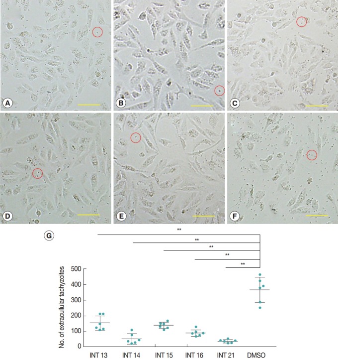
Fig. 2.

Sterculic acid and methyl esters inhibited the egress of tachyzoites from Vero cells. Representative pictures were presented to show that most of host cells remained intact after incubation with INT13 (A), INT14 (B), INT15 (C), INT16 (D), or INT21 (E) for 36 hr, with a small number of released tachyzoites, compared with numerous tachyzoites and a smaller number of intact host cells in the negative control group (F). Scale bar=100 μM. Extracellular tachyzoites are marked in red circles. (G) The number of released tachyzoites in the culture incubated with the test compounds was decreased relative to the DMSO control. Extracellular tachyzoites were counted in 6 random fields under the microscope per group. Data are presented as mean±SD. **P<0.01.
