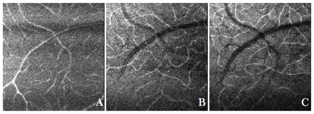
Fig. 3.
En-face images of the retina of a healthy volunteer with the focus set to the anterior layers retrieved from an AO-OCT data set. The entire data set can viewed in Visualization 3 (5.7MB, MOV) . A) Vasculature within the ganglion cell layer, B) vasculature within the inner plexiform layer and C) vasculature within outer plexiform layer.

