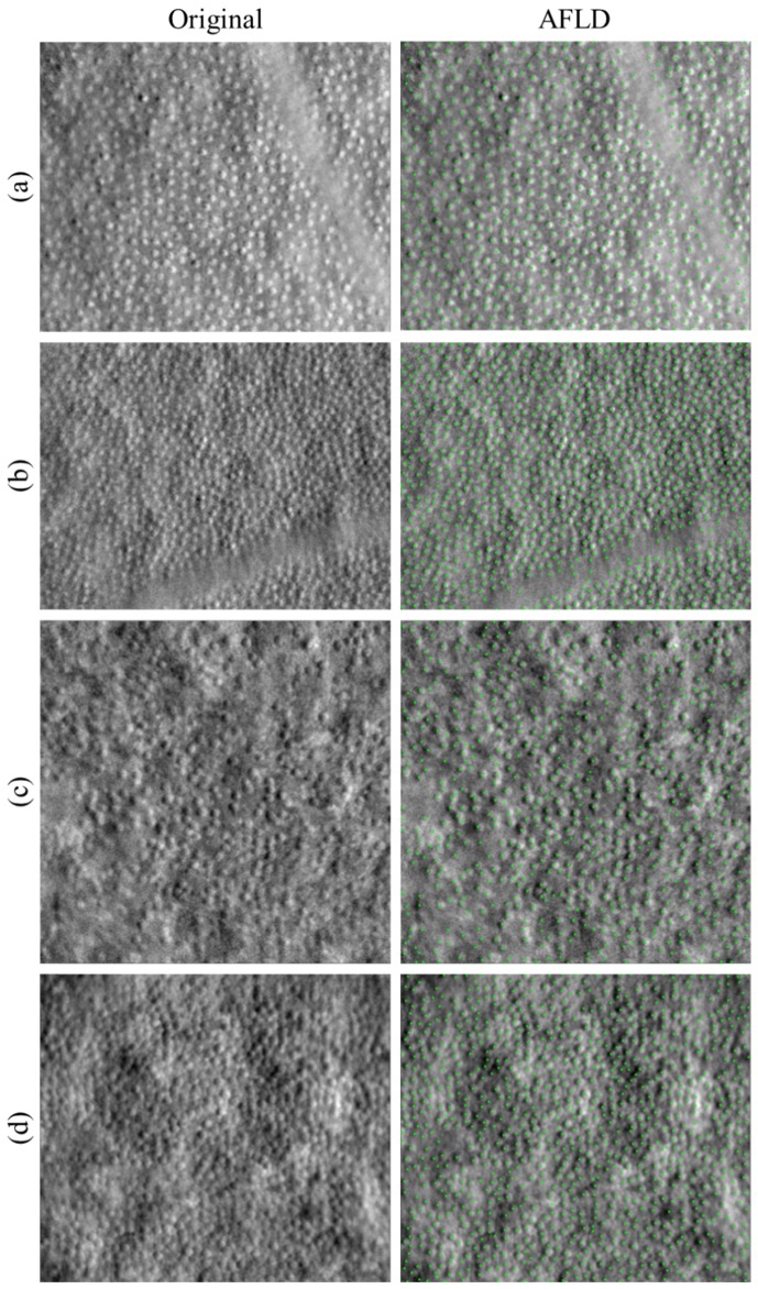
Fig. 9.
AFLD cone detection in split detector images of four subjects with photoreceptor pathology. Original images are shown in the left column, and images with automatically detected cones marked in green are shown in the right column. Subject pathologies are (a-b) oculocutaneous albinism and (c-d) achromatopsia.

