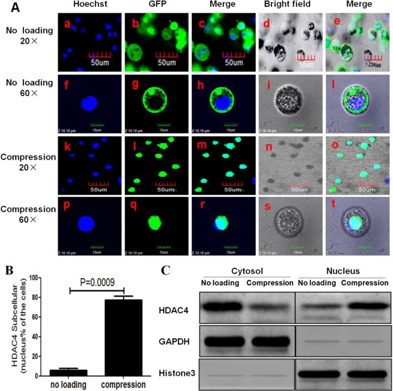
Figure 2.
Compression-induced HDAC4 nuclear import in chondrocytes. (A) Confocal microscopy showed that HDAC4 was mainly located in the nucleus of cells subjected to 3 h compression (A-k to t) when compared to unloaded cells (A-a to j). Blue indicated cell nuclei stained by Hoechst 33342, and green indicated the GFP-HDAC4 stain. (B) Percentage of green GFP-HDAC4 located in nucleus only. Approximately 300 cells from 3 independent experiments were scored. Data are expressed as means±SD (P=0.0009). (C) Cytoplasmic and nuclear lysates from the cells were separated by using the Nuclear Extract Kit and followed by western blot analysis with HDAC4 antibody. GAPDH and histone 3 are shown as loading controls for the cytoplasmic and nuclear fractions, respectively.

