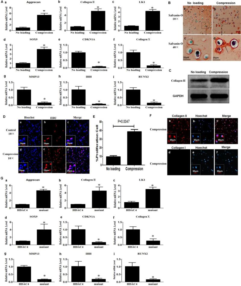
Figure 3.
Compression enhanced anabolism and proliferation, but inhibited differentiation markers in chondrocytes. (A) Real-time PCR was performed to detect the mRNA expression for aggrecan (A-a), type II collagen (A-b), LK1 (A-c), SOX9 (A-d) , CDKN1A (A-e), type X collagen (A-f), MMP-13 (A-g), Ihh (A-h) and Runx2 (A-i). The levels of mRNA in chondrocytes subjected to compression were compared with those in unloaded chondrocytes. Values are presented as mean±SD (n=3). *=P<0.05 versus the unloaded group. (B) Compression increased production of glycosaminoglycans. Safranin-O staining was performed to detect glycosaminoglycans at 48 h post-compression. The red staining indicated by arrow corresponds to glycosaminoglycans. (C) Compression increased type II collagen protein levels. Western blot analysis was performed with cell lysates collected 48 h post-compression with an antibody against type II collagen. (D,E) Compression stimulated chondrocyte proliferation. Forty-eight hours post-compression, cell proliferation was detected using an EdU-based cell proliferation assay kit. Values are presented as mean±SD (n=3). (F) Chondrocyte phenotype was confirmed by immunohistochemistry at 48 h post-compression. The cells were positive for type II, but not for type I collagen. (G) Transfection of HDAC4 S246/467/632A triple mutant increased the mRNA expression of aggrecan (G-a), type II collagen (G-b), LK1 (G-c), SOX9 (G-d), and decreased the mRNA express of CDKN1A (G-e), type X collagen (G-f), MMP-13 (G-g), Ihh (G-h) and Runx2 (G-i) compared with those transfected with Flag-HDAC4. Values are presented as mean±SD (n=3). *=P<0.05.

