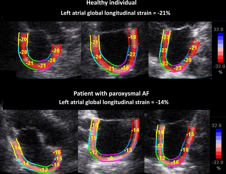
Figure 1.
Upper and lower panels demonstrating apical long-axis views (apical long-axis, two-chamber, and four-chamber views) with the left atrium as region of interest. Upper panels show a healthy individual with normal atrial deformation, while lower panels demonstrate reduced atrial deformation during atrial systole in a patient with PAF but in sinus rhythm during the study. AF, atrial fibrillation.

