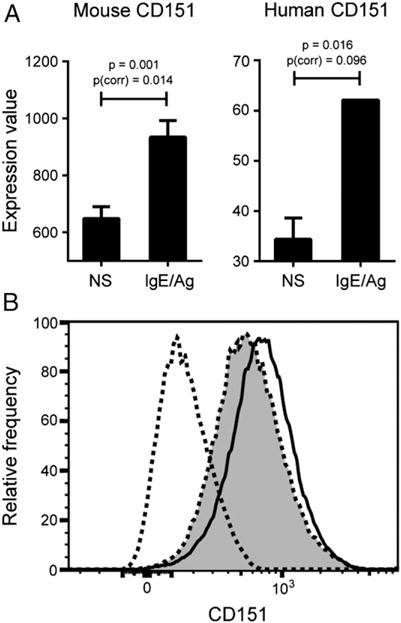
FIGURE 1.

IgE stimulation induces upregulation of tetraspanin CD151 expression by both mouse and human mast cells. (A) BMMCs from WT mice were either unstimulated or primed with 1 μg/ml DNP-IgE overnight, followed by 0.5 μg/ml DNP-HSA stimulation. Cells were collected 4 h after stimulation and subjected to gene expression microarray analysis using Illumina bead array technology. Nonstimulated mast cells (n = 3) were compared with IgE-stimulated mast cells (n = 3) using a t test with the Benjamini–Hochberg FDR multiple testing correction. This graph represents the average of three probe sets used to measure CD151 expression in the arrays (each individual probe showed statistical significance). Both raw t test p value and p value after FDR correction for multiple testing [p(corr)] are shown. Data for human mast cell CD151 expression are derived from GEO2R analysis of human immune cell transcriptomics study GSE3982 (National Center for Biotechnology Information’s GEO database). In this study, human cord blood–derived mast cells were either left unstimulated (n = 2) or stimulated with IgE for 2 h (n = 2). The p values shown are both unadjusted and adjusted by the Benjamini–Hochberg FDR correction [p(corr)]. p < 0.05 and p(corr) < 0.1 are deemed to be significant. (B) CD151 is upregulated in WT BMMCs at 4 h as measured by flow cytometry.
