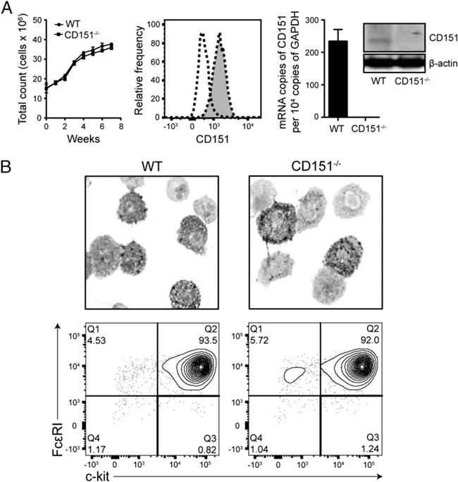
FIGURE 3.

CD151 deficiency does not alter basal phenotype of cultured BMMCs. (A) CD151 deficiency does not affect growth dynamics of mast cell culture. WT and CD151−/− BMMCs were cultured in IL-3–conditioned media for 8 wk and the total numbers of live cells in culture were counted at each time point indicated (left). Flow cytometry for CD151 expression on peritoneal mast cells (flow cytometry chart) (middle), qPCR detection of CD151 mRNA in WT and CD151−/− BMMCs (bar graph, right), and Western blot analysis of total-protein lysates for CD151 expression (Western blot, right) all confirm constitutive CD151 expression in WT mast cells. WT peritoneal mast cells are shown as gray-filled histogram with dotted line and negative control as transparent histogram with dotted line. In immunoblotting, actin was used as a loading control. (B) Five-week-old BMMCs from WT and CD151−/− mice were stained with toluidine blue and images were obtained with an original magnification of ×100. Flow cytometry analysis of FcεRI and c-Kit surface expression and purity of WT and CD151−/− BMMC cultures. All data are representative of three independent experiments. Data are represented as mean ± SEM. *p < 0.05.
