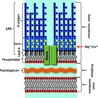
Figure 1.

Schematic representation of the Gram‐negative bacterial envelope, including the outer membrane (OM) with long “smooth” LPSs, core‐associated divalent cations, integral membrane proteins (in this case, a channel‐forming porin such as OmpF), and the inner phospholipid layer. The periplasm and inner membrane contain many proteins (not shown). Not to scale; the porins are approximately 5–6 nm high, the periplasm about 14 nm, and the inner membrane approximately 4 nm.
