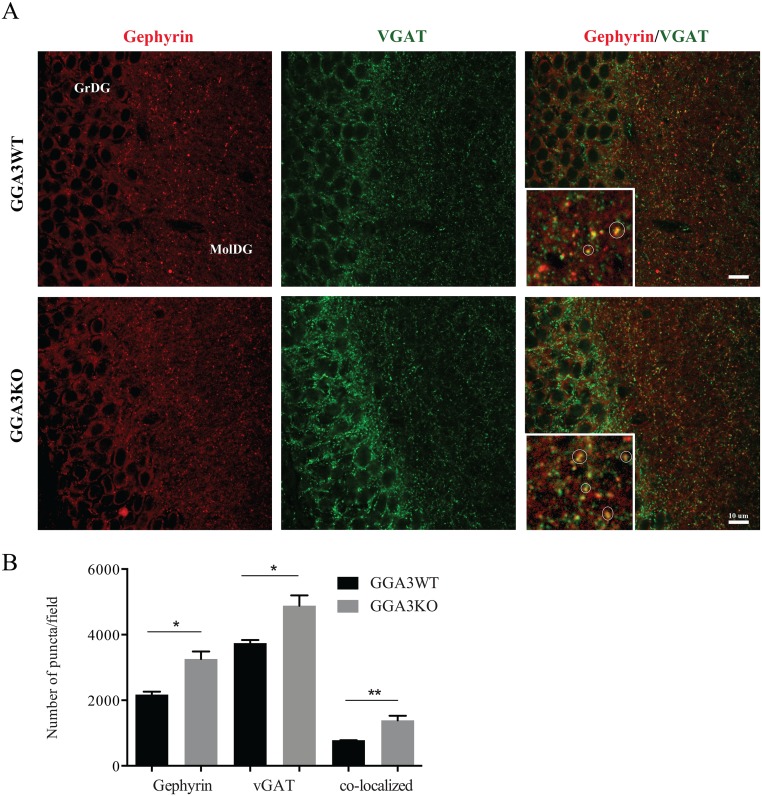
Fig 7. The number of inhibitory synapses is increased in the dentate gyrus of GGA3KO mice.
(A) 50 μm coronal brain slices were stained with pre- and post-synaptic marker antibodies (vGAT and gephyrin, respectively) and fluorescent conjugated secondary antibodies, followed by confocal microscopy. The numbers of vGAT (green, left panels), gephyrin (red, middle panels), and co-localizing puncta (vGAT/gephyrin, right panels) were quantified in the dentate gyrus of GGA3WT (upper panels) and GGA3KO mice (lower panels). A magnified image of the molecular granular layer of the dentate gyrus (MolDG) it is shown in the inset of the right panels, where the co-localizing puncta are highlighted within a circle. GGA3KO mice showed an increase in the presynaptic, postsynaptic, and co-localizing puncta. GrDG: Granular cell layer, MolDG: Molecular layer. Scale bar 10μm. (B) The graph represents mean ± SEM of the total number of puncta measured in 4 GGA3WT and 6 GGA3KO mice using Puncta Analyzer Plug-in. Two-four optical fields (116.4 x 114.4 x 5 μm) from each of two brain sections were analyzed for each mouse. The number of gephyrin, vGAT and co-localizing puncta were increased in GGA3KO mice compared to the wild-type littermates. Mann-Whitney test was used for statistical analysis, * p<0.05, **p = 0.0095.

