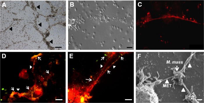
Fig 1. Microscopic examination of macrophage extracellular trap (MET) formation induced by M. mass infection.
Differentiated THP-1 macrophages were infected with M. mass R or CIP at a MOI of 5 for 24 hr, and stained by DNA-binding dye, TO-PRO-3 (Red) to visualize extracellular trap-like structure. (A) Microscopic examination of extracellular trap-like structures in M. mass R-infected THP-1 macrophages. Bar, 200μm. (B, C) Microscopic examination (B) and TO-PRO staining (C) of extracellular trap-like structures induced by M. mass R infection. Bar, 50μm. (D, E) Fluorescence micrographs of CFSE-stained M. mass R (Green) entrapped by METs. Bar, 20μm. (F) Scanning electron microscopy (SEM) images of METs induced by M. mass CIP. Bar, 1μm. Arrow: Mycobacteria. Arrow heads: Extracellular trap-like structures.

