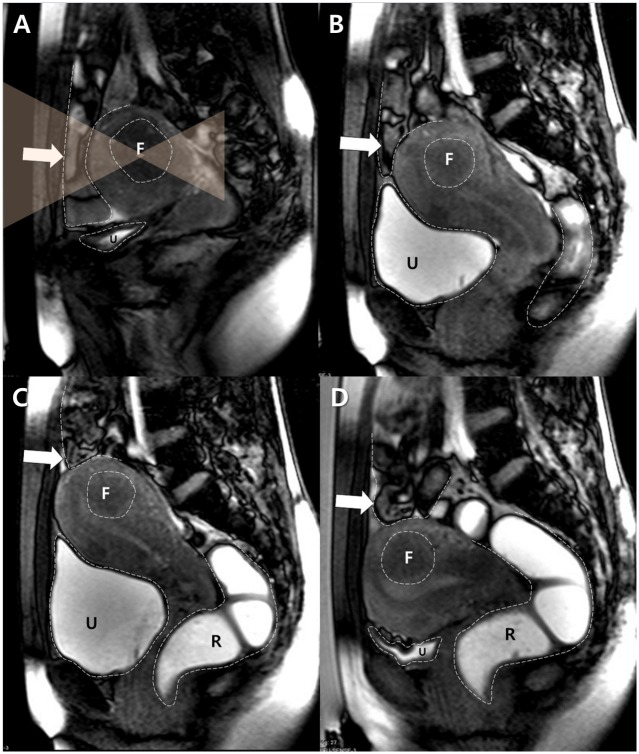
Fig 1. A typical example of the BRB maneuver attempted in the case of a 34 year-old woman with a fibroid in a forward-bent uterus.
A. A sagittal survey scan showed that the bowel loops (arrow) were interposed between the uterus and the anterior abdominal wall. F and U indicate the target fibroid and the urinary bladder respectively. Dotted lines delineate the margins of each structure, and transparent orange triangles represent the planned HIFU beam path. B. After filling with 500 mL of saline, the urinary bladder (U) was distended and displaced the uterus cranially. However, the interposed bowel loops (arrow) were still in the anticipated sonication path. F indicates the target fibroid. Dotted lines delineate the margins of each structure. C. The rectum (R) was filled with 150 mL of gel. The distended rectum pushed the uterine cervix and the uterus antero-cranially, which displaced the bowel loops (arrow) out of the anticipated sonication path. F indicates the target fibroid and U indicates the urinary bladder. Dotted lines delineate the margins of each structure. D. The uterus descended after drainage of the urinary bladder (U), although the previously-interposed bowel loops (arrow) remained out of the anticipated sonication path. After a successful BRB maneuver, MR-HIFU ablation was performed. F indicates the target fibroid and R indicates the rectum. Dotted lines delineate the margins of each structure.

