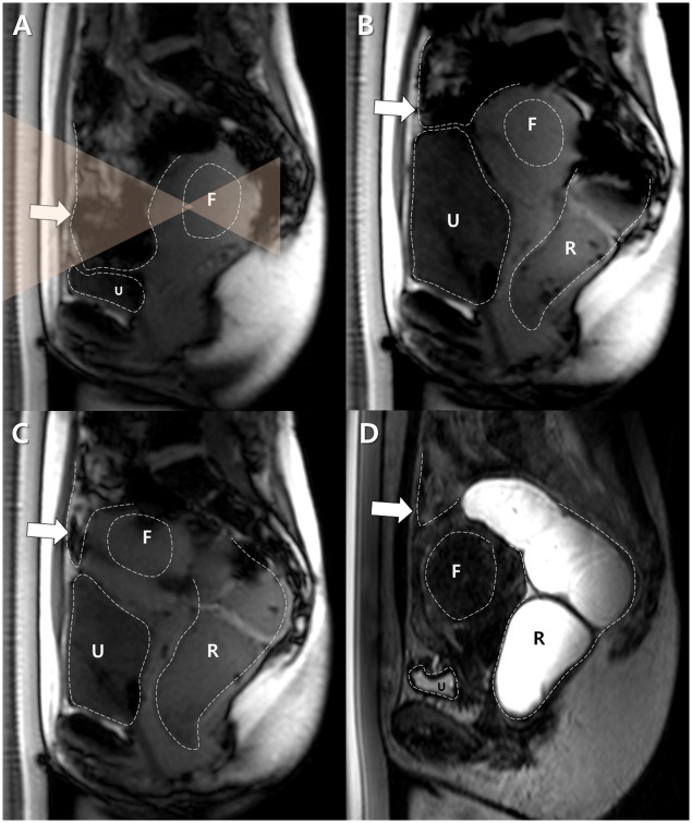
Fig 2. A successful BRB maneuver for a backward-bent uterus in a 43 year-old woman with a uterine fibroid.
A. A sagittal survey scan revealed the backward-bent uterus located in the deep pelvic cavity and the interposed bowel loops. The size of the uterus was 90 mm in its largest dimension. F and U indicate the target fibroid and the urinary bladder, respectively. Dotted lines delineate the margins of each structure, and transparent orange triangles represent the planned HIFU beam path. B. The urinary bladder (U) was filled with 500 mL of saline and then the rectum (R) was filled with 100 mL of gel. The uterus was shifted antero-crainally. However, the bowel loops (arrow) continued to block the target fibroid (F). Dotted lines delineate the margins of each structure. C. The urinary bladder (U) was partially emptied by draining 100 mL of urine and the rectum (R) was filled further with 100 mL of gel. The target fibroid (F) was shifted anteriorly, but the bowel loops (arrow) were still in the anticipated sonication path. Dotted lines delineate the margins of each structure. D. After fully emptying the urinary bladder (U), the uterus was moved antero-caudally close to the abdominal wall, and the bowel loops (arrow) were displaced completely out of the sonication path. After a successful BRB maneuver, MR-HIFU ablation was performed. F indicates the target fibroid and R indicates the rectum. Dotted lines delineate the margins of each structure.

