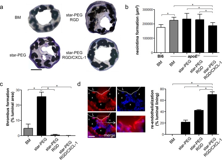
Fig 3. RGD/CXCL1-biofunctionalized stents reduce neointima formation.
(a-d) Carotid arteries of C57Bl/6 (n = 6) or apoE-/- mice were analyzed one week after implantation of bare metal nitinol-stents (BM, n = 4), star-PEG-coated nitinol-stents (n = 5) or star-PEG-coated stents bio-functionalized with RGD (n = 9) or RGD/CXCL1 (n = 8). (a) Representative Giemsa-stained sections (scale bars, 200μm) from apoE-/- mice. Quantification of in-stent intima formation (b) and thrombus formation (c). (d) Representative images of vWF-staining (red); cell nuclei were counterstained by DAPI (L, lumen; x, stent struts; scale bar, 50μm); quantification of re-endothelialization. *P<0.05. Kruskal-Wallis test with Dunn’s post-test.

