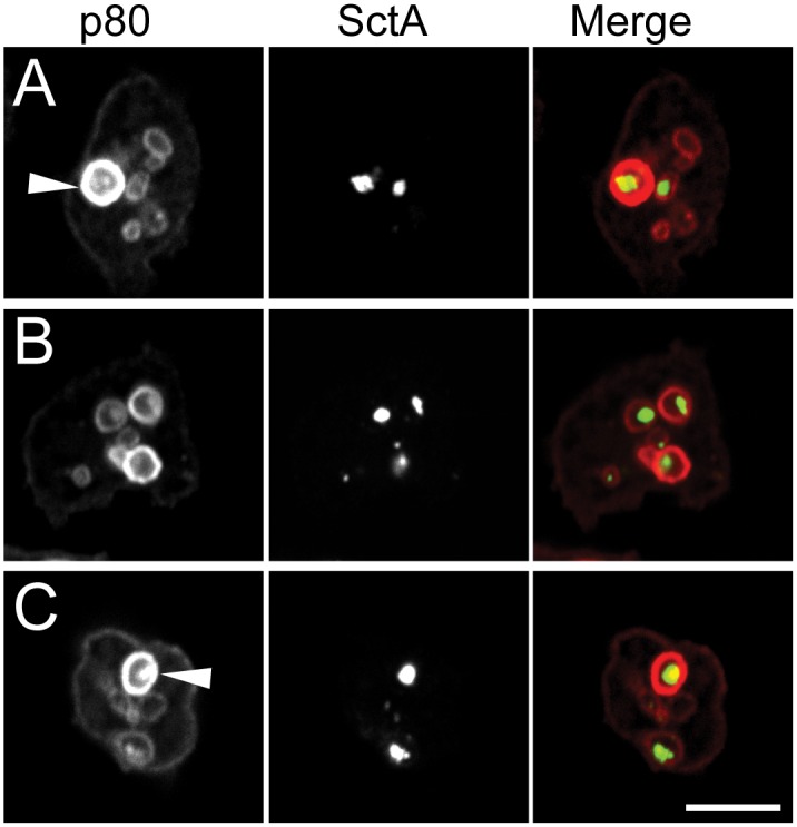
Fig 5. SctA-positive structures are detected in endocytic compartments.

Confocal images of three D. discoideum cells, labeled for SctA protein (middle column, green) and for endosomal p80 (left column, red) and merged images (right column) are shown. SctA-positive structures were detected both in large p80-rich post-lysosomes (arrowheads) and in p80-low endosomes. Bar: 10 μm.
