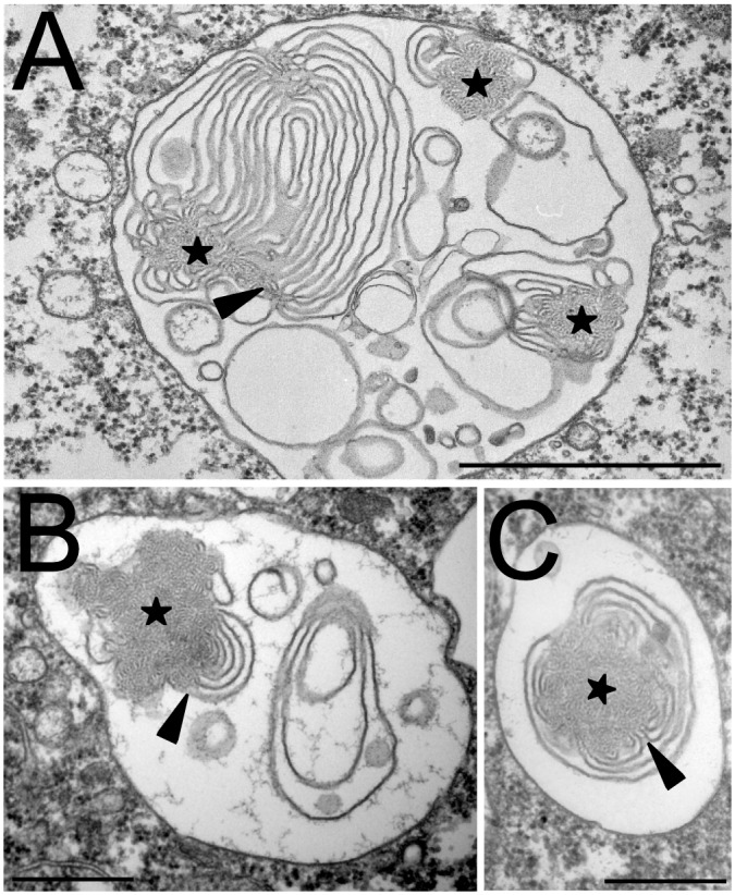
Fig 7. Intraendosomal membranes association with pycnosomes.

D. discoideum cells were treated for 2h with U18666A to induce the formation of intraluminal membranes in the endocytic compartments, then fixed and processed for electron microscopy. Pycnosomes (stars) often appeared continuous with internal membranes. Arrowheads point to regions where the continuity between pycnosomal material and endosomal membranes was most apparent. Bar: 500 nm.
