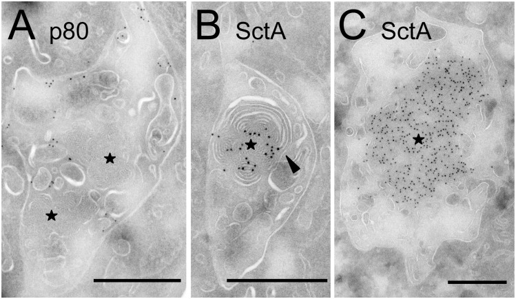
Fig 8. SctA-positive material appears continuous with intraendosomal membranes.
D. discoideum cells were treated for 2h with U18666A, then fixed and processed for immuno-electron microscopy. (A) In sections labeled with the H161 anti-p80 antibody, the p80 protein was seen associated with the limiting endosomal membrane as well as with internal membranes, but was not detected in pycnosomes (stars). (B-C) SctA-positive structures with the dense morphology of pycnosomes were also detected, and often appeared continuous with intraluminal membranes (arrowhead). Bar: 500 nm.

