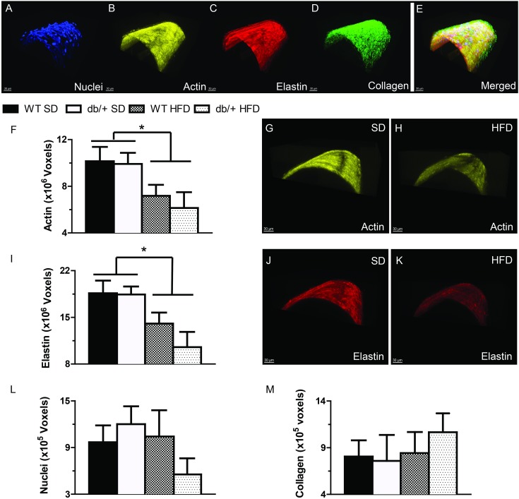
Fig 9. Effect of maternal environment and offspring diet on the morphological characteristics of mesenteric resistance arteries from adult (31 week-old) wild type (WT) male offspring of WT-control or Leprdb/+ (db/+) dams.
(A-E) Representative confocal images of mesenteric resistance arteries showing (A) nuclei; (B) F-actin; (C) elastin; (D) collagen; and (E) merged image. (F-H) Group data and representative images showing that feeding a high-fat diet was associated with a significant reduction in arterial F-actin content. (I-K) Group data and representative images showing that feeding a high-fat diet was associated with a significant reduction in arterial elastin content. (L) Vascular smooth muscle cell number, represented by nuclei contained within the medial layer of mesenteric arteries. (M) The amount of collagen contained in the wall of mesenteric arteries. Data are means ± SEM of n = 5–7 number of animals (vessels) per treatment group combination. *P<0.05. SD, standard diet; HFD, high fat diet.

