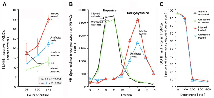
Fig 2. Effect of deferiprone in HIV-infected primary cells: Apoptotic DNA fragmentation, deoxyhypusyl-eIF5A accumulation, and DOHH activity.
Apoptotic DNA fragmentation (Panel A) was detected by terminal deoxynucleotide transferase dUTP nick end labeling (TUNEL) of single-donor peripheral blood mononuclear cells, maintained in short-term non-replenished cultures as described [43]. Cells were left uninfected and/or untreated; or infected immediately after plating with clinical HIV-1 isolate [43] and 12 hours after infection, treated with 200 μM deferiprone. In these single-donor cultures, metabolic labeling with (1,8-3H)spermidine was performed for quantification of the 3H-labelled deoxyhypusyl substrate and of the 3H-labelled hypusine product of cellular DOHH (Panel B), followed by acid hydrolysis, ion-exchange-based separation of the 3H-labeled amino acids, and fractionation as described [28,43,44,124]; the x-axis of the graph shows 8 of the 16 fractions from a representative experiment. The dose-effect curve for inhibition of cellular DOHH by increasing deferiprone concentrations in short-term non-replenished single-donor cultures (Panel C) was calculated from the accumulation of its 3H-labeled substrate and the decrease of its 3H-labeled product; error bars are at the size of the graph symbols and not shown. Black, HIV-infected untreated cells; green, uninfected untreated cells; blue, uninfected deferiprone-treated cells; red, HIV-infected deferiprone-treated cells; large triangles, HIV-infected cells (red) or uninfected cells (blue) treated with 200 μM deferiprone.

