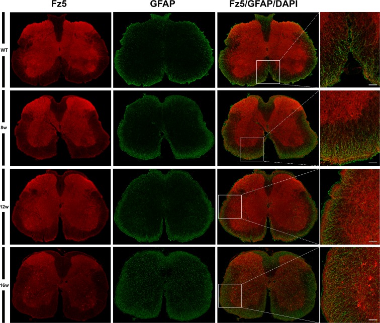
Fig 6. Fz5 receptor was localized in astrocytes.
This figure shows representative images obtained from the microscopic evaluation of sections processed by double immunohistochemistry to visualize Fz5 in astrocytes (GFAP). Fz5 receptor was located in astrocytes both WT and ALS transgenic mice, mainly in contacting pial surface regions, but no changes were observed during disease progression. The analysis was performed in WT and ALS mice spinal cords at 8w, 12w and 16w. The squares in the images showing the entire spinal cord sections represent the areas where the corresponding higher magnification micrographs were obtained. Scale bars = 50 μm.

