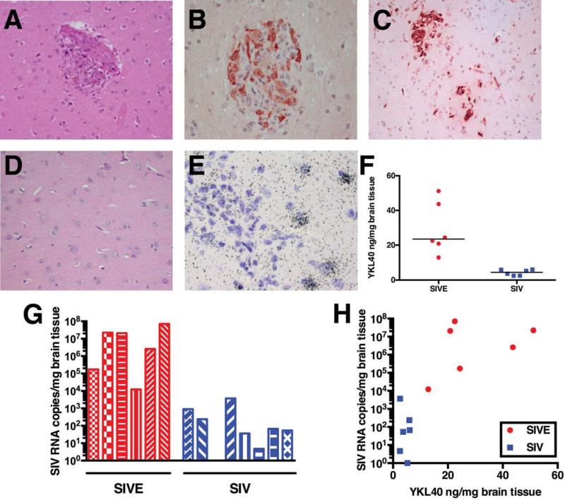
Fig. 1. Macaques with SIV encephalitis show SIV-infected microglial nodules and increased SIV viral load and YKL40 expression in frontal cortical tissue.
Macaques were defined as having SIV encephalitis (SIVE) by histological examination of brain sections. Hematoxylin and eosin (H&E) stains show the presence of perivascular cuffing, a microglial nodule and multinuclear giant cells (a). Many cells in microglial nodules stain with an antibody against the SIV envelope protein (b). Microglial nodules and macrophage infiltrates can be observed with CD68 immunostains (c). SIV-infected macaques without encephalitis show no infiltrate (H&E) (d). In situ hybridization for YKL40 shows cells expressing YKL40 RNA surrounding a microglial nodule (e). YKL40 concentrations in frontal cortical tissue extracts of macaques with SIVE (red) are significantly elevated compared to SIV-infected macaques without encephalitis (blue), P = 0.002 (f). SIV RNA viral loads in frontal cortical tissue from macaques with SIVE (red) are higher than SIV-infected macaques without encephalitis (blue), P = 0.0007 (g). Correlation between frontal cortical SIV RNA viral load and frontal cortical YKL40 levels (h).

