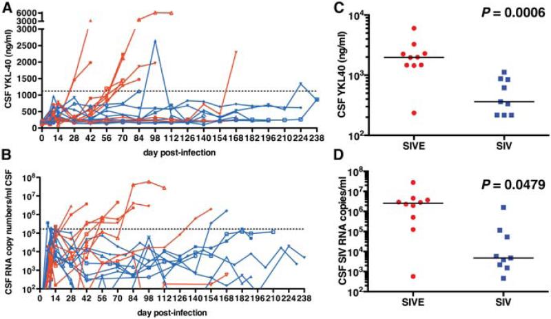
Fig. 2. Macaques that develop encephalitis have increased CSF YKL40 and SIV RNA concentrations.
Preserved CSF samples obtained from 19 macaques from prior studies were analyzed for YKL40 concentrations (a) and SIV viral load (b). Macaques that were determined to have presence of SIVE based on post-mortem histological findings are shown in red while SIV-infected non-encephalitic macaques are depicted in blue. The dashed lines (a and b) represent the concentration determined statistically likely to be associated with encephalitis, 1122 ng/ml for CSF YKL40 concentrations and 1.65 × 10⁵ copies/ml CSF for SIV CSF viral load. Necropsy analyses of terminally ill SIV-infected non-encephalitic macaques (blue) show a significantly lower CSF YKL40, P = 0.0006 (c) and CSF SIV viral load, P = 0.0479 (d) than macaques with SIVE. The black lines represent the median.

