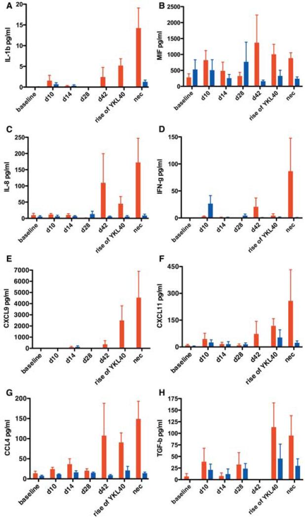
Fig. 6. The majority of elevated neuroimmune markers became elevated as encephalitis developed and were associated with macrophage recruitment and activation.
Multiplex quantitation of 31 cytokines present in the CSF was performed on samples from baseline (d0), acute infection (d10 and d14), asymptomatic infection (d28 and d42), development of encephalitis (rise of YKL40), and at necropsy (nec). IL-1β (a), MIF (b), IL-8 (c), IFN-γ (d), CXCL9 (e), CXCL11 (f), CCL4 (g), TGF-β (h) were elevated when encephalitis (red) developed or shortly before. SIV-infected non-encephalitic macaques (blue) show little elevation of these markers. Bars represent median concentrations of the indicated cytokine.

