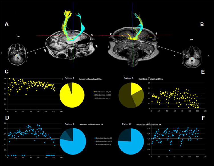
Fig. 3.

In order to determine FA of the CSTs, spheres of 3 mm (123 voxels) were created symmetrically in both tracts, their middle centered on the CS fibers as visualized in a transversal plane passing through the middle cerebellar peduncle (see picture in transversal plane). The number of voxels presenting a main z direction was then counted, and the value of the FA in z direction reported for the 123 voxels in each sphere. A determinist tracking was then made from the spheres. Only the fibers with FA > 0.15 were considered for this tracking and a deviation angular of less than 508 was required. The upper panels (A and B) represent the tracking for children 1 and 2 respectively. The lower panel delineates the fractional anisotropy of the fibers for right (yellow) and left (blue) CST respectively in both children. Fig. 4a: Child 2/at same q.
