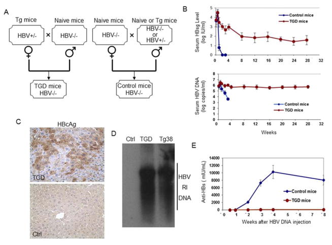
Figure 1. HBV persists in TGD mice but not in control mice.
(A) Procedures for the generation of TGD mice and control mice. HBV+/− mice were hemizygous Tg05 or Tg38 HBV transgenic mice. (B) Analysis of HBV persistence in control and TGD mice. The sera of 12 control mice and 13 TGD mice injected with 20 μg 1.3mer HBV DNA were collected at the time points indicated and analyzed for HBsAg by ELISA (upper panel) and HBV DNA by real-time PCR (lower panel). (C) Immunostaining of HBV core protein in the liver of TGD mice (upper panel) and control mice (lower panel). (D) Southern-blot analysis of HBV replicative intermediate (RI) DNA in the mouse liver. The liver tissues of control (Ctrl) and TGD mice were isolated at three months after HBV DNA injection for the analysis. The liver tissue of a 3-month old Tg38 mouse was also analyzed to serve as the control. (E) Analysis of anti-HBsAg antibodies in mouse sera by ELISA. See also Figure S1.

