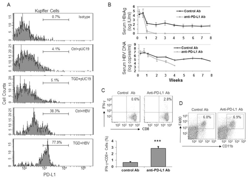
Figure 3. Enhanced expression of PD-L1 in Kupffer cells is critical for HBV persistence in TGD mice.
(A) Analysis of PD-L1 expression in Kupffer cells isolated from control (Ctrl) and TGD mice that were injected with pU19 or HBV DNA. The isotype antibody was used as the negative control to stain Kupffer cells isolated from control mice that were injected with pUC19. (B) Analysis of the effect of the anti-PD-L1 antibody on HBV persistence in TGD mice. TGD mice were treated with either the control antibody or the anti-PD-L1 antibody. The serum samples were collected at the time points indicated for HBsAg (upper panel) and HBV DNA (lower panel) analysis. Three different animals were used for each group and the results represented the mean. (C) Analysis of HBV-specific CD8+ T cells. TGD mice treated with either the control antibody or the anti-PD-L1 antibody were analyzed by flow cytometry using the procedures described in the Figure 2 legend. Representative flow cytometry results are show on the top. The results shown in the histogram represent the mean of five different mice in each group. ***, p<0.005. (D) Analysis of the effect of the anti-PD-L1 antibody on Kupffer cells. Kupffer cells were isolated from TGD mice two days after the injection of the control antibody or the anti-PD-L1 antibody and analyzed by flow cytometry.

