
Figure 3. Genetic removal of VG3-ACs reduces inhibition of SbC-RGCs in a contrast- and size-selective manner.
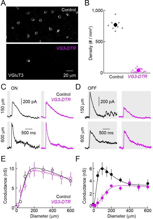
(A) Representative z-axis projections of confocal image stacks of retinal whole mounts stained for VGluT3 in control (top) and VG3-DTR mice (bottom) one week after diphtheria toxin injections. (B) Summary data of VG3-AC density in control (black) VG3-DTR (purple) retinas. Dots show data from individual retinas (control, n = 8; VG3-DTR, n = 18, p < 10−20) and circles (errorbars) represent mean (± SEM). (C, D) Representative IPSCs in SbC-RGCs elicited by light increments (C, ON) and decrements (D, OFF) in small (150 μm diameter, top) or large (600 μm diameter, bottom) circles recorded in control (left, black) and VG3-DTR (right, purple) retinas. (E, F) Summary plots (mean ± SEM) comparing inhibitory synaptic conductances in SbC-RGCs of control (n = 5, black) and VG3-DTR (n = 4, purple) retinas elicited by ON (E) and OFF (F) stimuli of different sizes (i.e. circle diameters). Inhibitory conductances elicited by small and large ON stimuli are unaffected by deletion of VG3-ACs (e.g. p > 0.7 for control vs. VG3-DTR at 150 μm and 600 μm). Inhibitory conductances activated small (p < 0.01 for control vs. VG3-DTR at 150 μm) but not large (p > 0.5 for control vs. VG3-DTR at 600 μm) OFF stimuli are reduced by removal of VG3-ACs. See also Figure S3.