
Fig. 2. The DLO/AI innervates the lateral and ventral striatum and BLA.
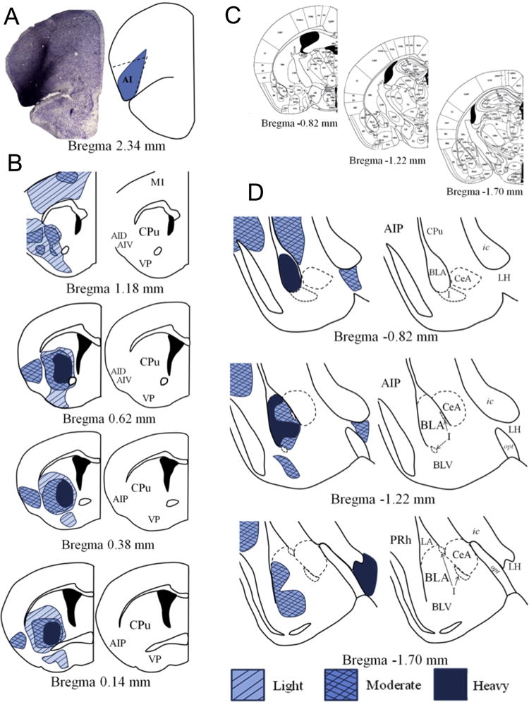
(a) A representative infusion of BDA into the DLO/AI and a rendering of the targeted area are shown. (b) BDA infusions into the DLO/AI illuminate heavy innervation of the ventral and lateral striatum, and reveal innervation of the posterior AI that is maintained along the rostrocaudal axis. (c) Coronal amygdala sections from (16) correspond to the magnified depictions shown in (d). (d) The DLO/AI sends heavy projections to the anterior BLA. Projections are lighter in the posterior BLA, and preferentially terminate along the lateral wall. Innervation is also noted in the ventral BLA, as well as the posterior AI and the PRh. Abbreviations not defined in Fig. 1: AID-dorsal agranular insular cortex; BLV-basolateral amygdala, ventral part; LH-lateral hypothalamus; M1-primary motor cortex; PRh-perirhinal cortex; VP-ventral pallidum.