
Fig. 3. Photomicrographs show representative BDA staining and demonstrate innervation of the striatum and PRh and BLA by the DLO/AI.
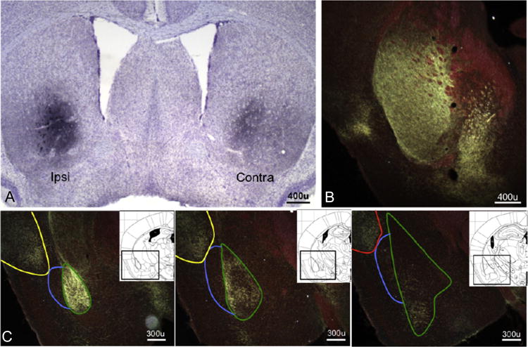
(a) Projections from the DLO/AI to the striatum are bihemispheric, but labeling is heaviest in the hemisphere ipsilateral to the infusion site, and (b) heavy labeling is noted in the posterior caudate. (c) Representative images of DLO/AI innervation of the BLA, posterior AI, and PRh (rostral to caudal); note avoidance the dorsal endopiriform nucleus. Green outline-BLA; blue outline-DEn; yellow outline-posterior AI; red outline-PRh. Inset: Corresponding images from (16), with regions outlined in black.