Figure 3.
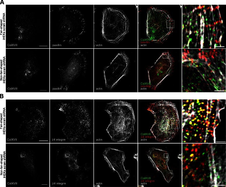
Localization of ColXVII, β4 integrin, paxillin, and actin in control keratinocytes. Control keratinocytes were triple stained with rhodamine-conjugated phalloidin and antibodies against ColXVII in combination with paxillin (A) or β4 integrin (B). Fourth column shows overlays of 3 stains. Boxed areas are shown at higher magnification in fifth column for each set of micrographs. Top row of each panel is fan-shaped cell, bottom row non-fan-shaped. Scale bars, 10 μm (left column) and 2 μm (right column).

