Figure 5.
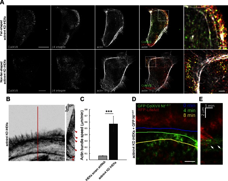
Actinin4 regulates ColXVII and β4 integrin localization and restricts actin dynamics in live keratinocytes. A) Actinin4 KD keratinocytes were triple stained with rhodamine-conjugated phalloidin and antibodies against ColXVII and β4 integrin. Fourth column shows overlays of 3 stains. Boxed areas are shown at higher magnification in fifth column. Scale bars, 10 μm (left column) and 2 μm (right column). B) Actin bundle retrograde movement was recorded in RFP-tagged LifeAct expressing actinin4 KD keratinocytes (n = 8). Kymograph in right panel was generated along line indicated in panel on left. Red arrowheads indicate bundle movement. C) Quantification of actin bundle retrograde movement. Values are means ± sem. ***P < 0.001. Mann-Whitney U test. D) High-magnification image taken from time-lapse fluorescent microscopic analyses of actinin4 KD keratinocyte expressing both GFP-tagged ColXVII Nt1–517 and RFP-tagged LifeAct. Actin bundle was followed over time; its position is marked by colored lines at indicated time points. Scale bar, 2 μm. E) Kymograph generated from (D). White arrow indicates an actin bundle that moves unimpeded over ColXVII puncta.

