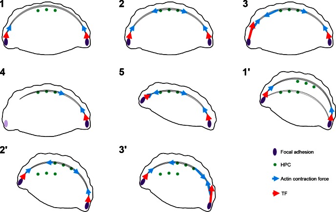
Figure 7.
Model of role of HPCs, focal adhesions, and actin in generation of TFs and cell movement (see Discussion for details). In (1), actin bundles undergo retrograde movement until they encounter HPCs (2). Because HPCs anchor actin bundles to the substrate, contraction can be localized (blue arrows) to one flank, resulting in larger TFs at the left flank vs. right [TF magnitudes represented by large and small red arrows in (3)]. In (4), detachment of actin bundles from focal adhesions at the left flank allows the cell to pivot, as shown in (5). In (1′), HPCs disengage from actin bundles and new HPCs reassemble along the lamella. In (2′), these new complexes reengage with actin moving in a retrograde fashion. In (3′), TFs predominate at the right flank due to localized contraction of the actin cytoskeleton to allow the cell to move stepwise over its substrate.

