
Figure 5.8.7.
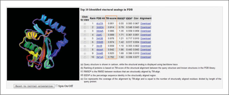
Ten PDB structures close to the target. The structure of the first I-TASSER model (model 1, shown in rainbow cartoon) is superimposed on the analogous structures from the PDB (shown in medium-purple backbone trace). The structural similarity between the target model and the 10 closest proteins are ranked by TM-scores, which are highlighted in the orange box. The coordinate file of the superimposed structures can be downloaded through the Download link for local visualization. In this example, there are multiple analogous structures from the PDB that have a high TM-score (>0.9), including 4co7A, 3m95A, and 3dowA. However, it is also possible that no similar structures can be found in the PDB; this usually indicates that the target protein is a new-fold protein or the fold by I-TASSER prediction is not correct.