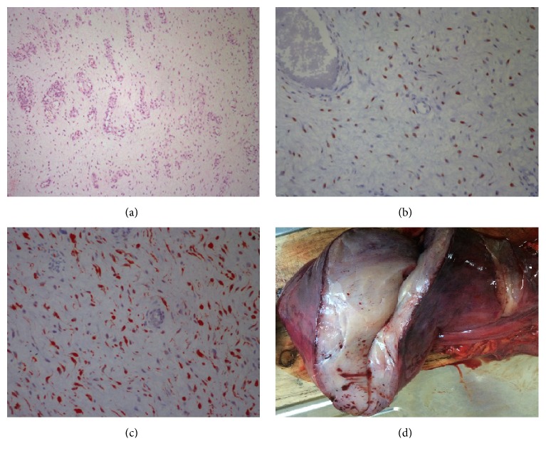
Figure 3.
Histopathologic examination of the tumor. (a) Spindled and stellate-shaped cells in a myxoid and richly vascular background (H&E ×100). (b) Estrogen receptor immunoreactivity in aggressive angiomyxoma (H&E ×400). (c) Diffuse desmin immunoreactivity in aggressive angiomyxoma (H&E ×400). (d) Macroscopic imaging of the tumor.

