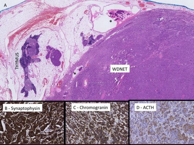
Figure 2.

(A) H&E of residual thymic tissue and well-differentiated neuroendocrine tumour (WDNET) with foci of lymphatic space invasion (*). The mitotic count is 1 per 10 HPFs and there is no necrosis. Immunohistochemical studies show positive immunoreactivity with synaptophysin (B), chromogranin (C) and ACTH (D).

 This work is licensed under a