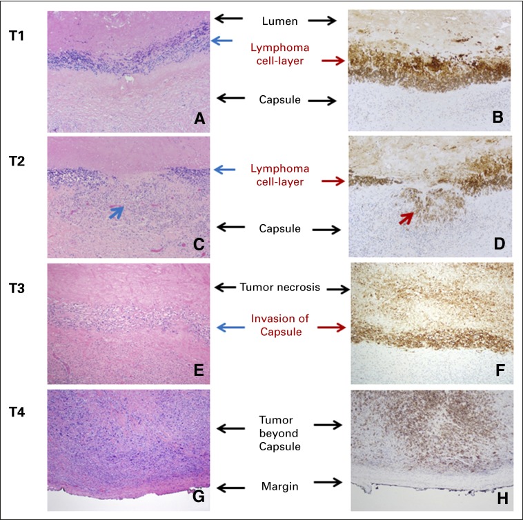
Fig 1.
Pathologic T staging. (A and B) T1: lymphoma cells confined to the effusion or a layer on the luminal side of the capsule; (C and D) T2: lymphoma cells superficially infiltrate the luminal side of the capsule. Arrows indicate the areas of invasion; (E and F) T3: clusters or sheets of lymphoma cells infiltrate into the thickness of the capsule; and (G and H) T4: lymphoma cells infiltrating beyond the capsule, into the adjacent soft tissue or breast parenchyma. Left column, hematoxylin and eosin stain; right column, CD30 immunohistochemistry; magnification, ×100.

