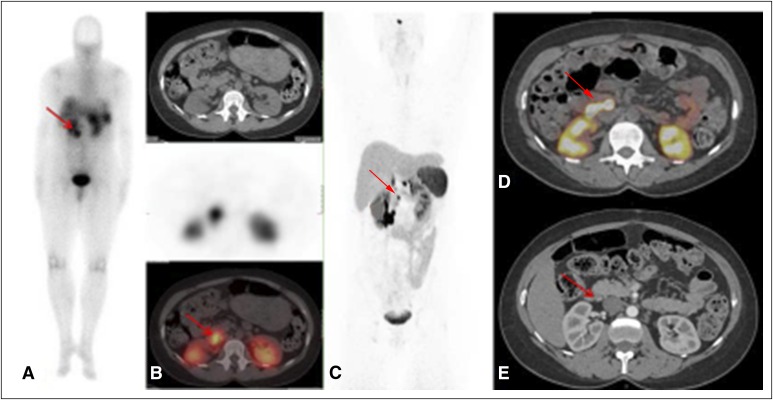
Fig A1.
A 46-year-old patient with multiple endocrine neoplasia type 1 and known pancreatic and duodenal lesions who had lymph nodes not previously known that were detected by using 68Ga-DOTATATE positron emission tomography (PET)/computed tomography (CT) imaging. (A) 111In-Pentetreotide scan (planar) shows a unique unclear prerenal uptake. (B, top) Axial CT, (middle) 111In-pentetreotide axial slice, and (bottom) fused single-photo emission CT/CT showing unclear pathologic uptake (red arrow). (C) 68Ga-DOTATATE PET maximum-intensity projection image shows retropancreatic lymph node (red arrow) and duodenal and pancreatic lesions. (D) 68Ga-DOTATATE Q:17 PET/CT image shows the retropancreatic and periduodenal lymph node (maximum standardized uptake value, 96; red arrow). (E) Arterial phase CT shows corresponding lymph node (red arrow) that was read as a subcentimeter indeterminate lymph node.

