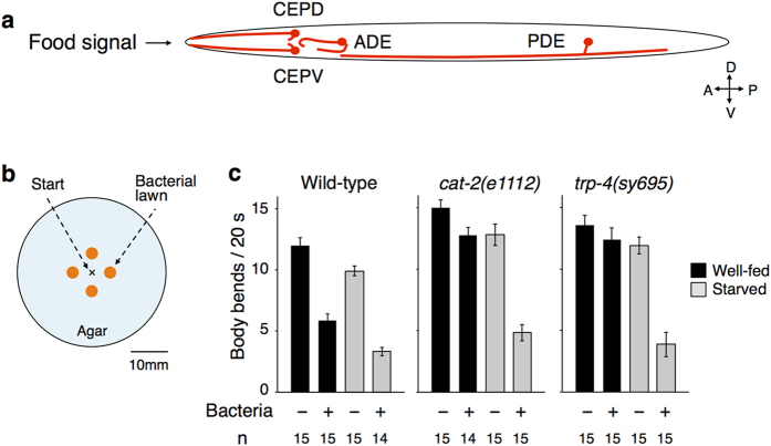
Figure 1. DAergic neurons mediate food-dependent slowing behaviour.
(a) A schematic diagram of arrangement of the four DAergic neuron pairs. Note that this is the top view of an animal on an agar surface. (b) An arrangement of patches of bacterial lawns on an agar surface. Similar experimental condition was recently reported by Hardaway et al.38. (c) Food-dependent slowing response of wild-type and mutant animals. Well-fed animals (black bars) or animals starved for 30 min (grey bars) were transferred to the centre position of the assay plate shown in b and the bending numbers were scored for 20 s after the food entry. The numbers of animals used in each condition are shown at the bottom.

