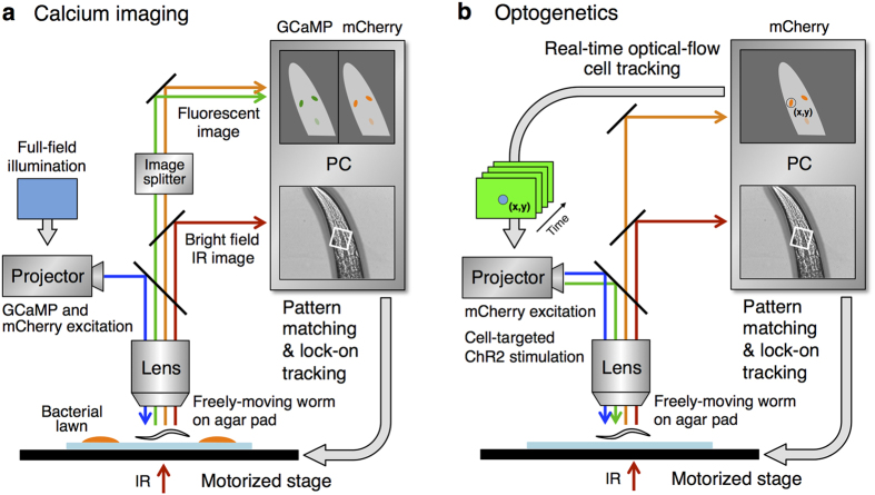
Figure 2. Schematic drawing of the OSaCaBeN system.
Setups for calcium imaging (a) and optogenetic analysis (b) are shown. The motorized stage is controlled to lock-on a part of the animal’s body at the centre of the view field. In (a), blue light is illuminated to the full view field to excite GCaMP6f and mCherry in all of the DAergic neurons. In (b), green light for mCherry is illuminated to the full view field to monitor the positions of all of the DAergic neurons, and blue light is illuminated to one of the DAergic neurons to stimulate ChR2 when necessary. The position of the blue light illumination is updated in real time because the target neuron moves in the view field due to the limitation of tracking accuracy. An IR light was used to acquire bright field images for pattern matching and tracking.

