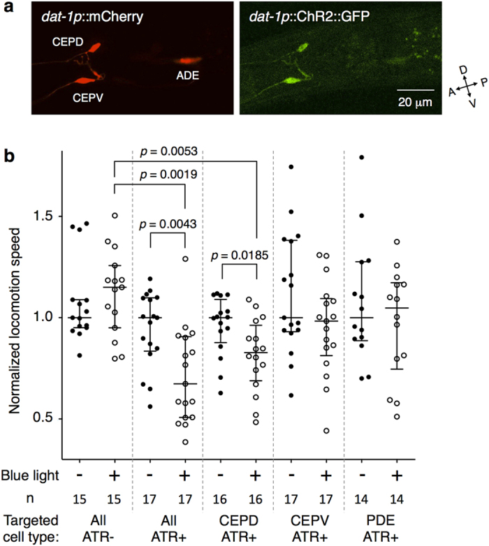
Figure 4. Activation of CEPD neuron pairs is mainly responsible for the slowing behaviour.

(a) Expressions of mCherry and ChR2::GFP were at similar levels between CEPD and CEPV but was lower in ADE. (b) Comparison of the effects of optogenetic stimulation on slowing. When all of the dopaminergic neurons or only CEPD were illuminated, but not CEPV or PDE, significant slowing occurred. A Mann-Whitney test was used to compare the normalized locomotion speeds before (−) and during (+) the blue light illumination within each targeted cell type, while a Kruskal-Wallis test with a post-hoc Steel-Dwass test was used to compare cell types during illumination. Details of the statistical analyses are shown in Supplementary Table S4.
